Single-Cell RNA-seq Experimental Workflow
A left-to-right scRNA-seq pipeline figure — tissue dissociation, droplet capture, library preparation, sequencing and clustering — with each stage labeled for methods sections.
更新日 2026-09-24
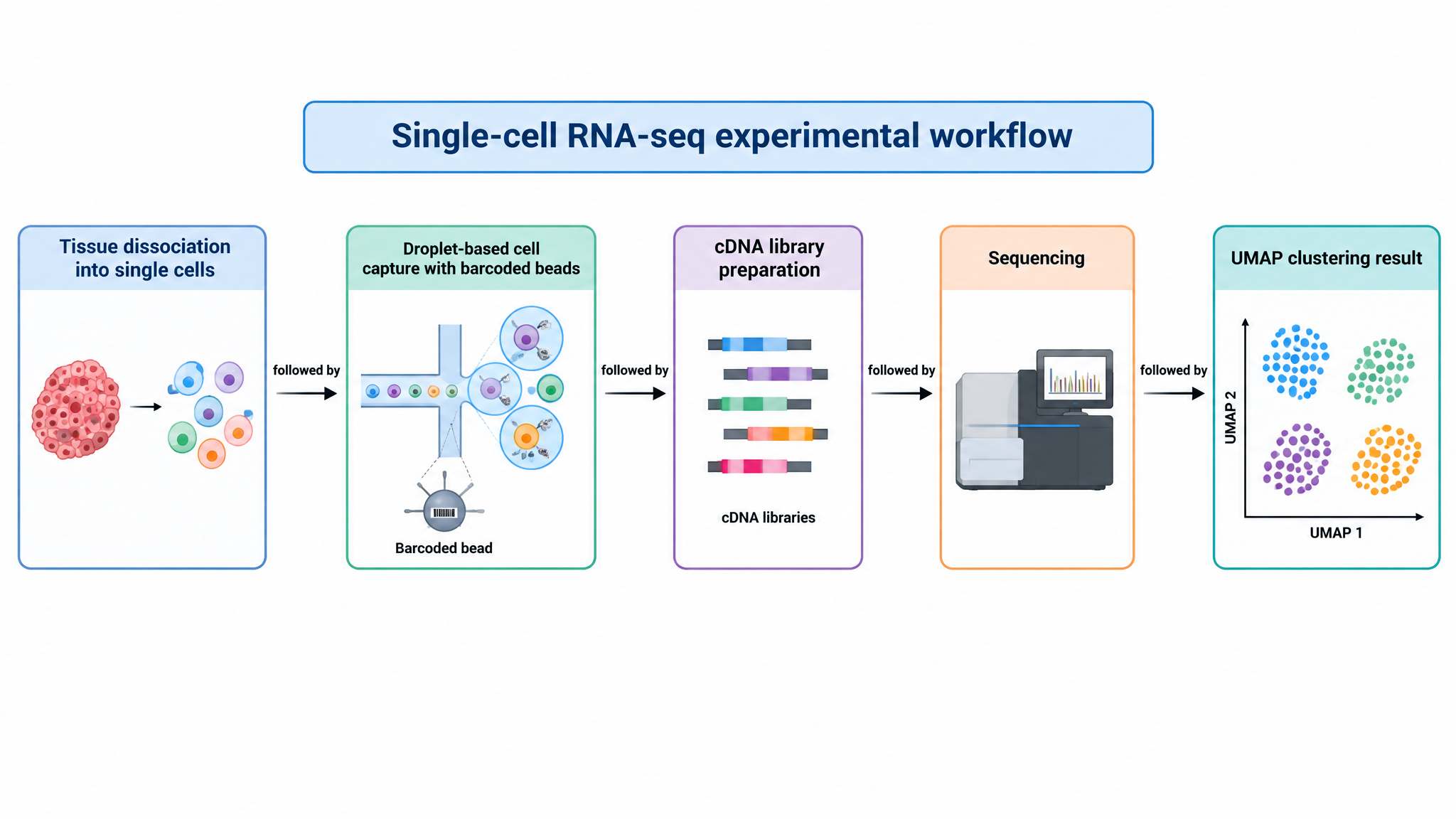
A single-cell RNA-seq experimental workflow diagram, left to right: tissue dissociation into single cells, droplet-based cell capture with barcoded beads, cDNA library preparation, sequencing, and a UMAP clustering result. Label each stage with its method name.
このテンプレートとは?
Workflow figures map a protocol end to end so reviewers can audit the experimental design at a glance. For omics studies they usually sit in methods or as figure 1 panel A.
適した場面
- Figure 1 study-design panels
- Methods-section protocol overviews
- Grant proposal technical approaches
- Core facility and training documentation
自分の図にする方法
Open the template in the studio
Generate the five-stage pipeline, then rename stages to your protocol.
Match your platform
Say "replace droplet capture with plate-based Smart-seq" or "add a nuclei isolation step" to reflect your actual method.
Align with your readout
Swap the UMAP for the analysis you actually show — trajectory, cell-cell communication, differential expression.
よくある質問
Can the same layout work for other omics?
Yes. The five-stage spine transfers to ATAC-seq, spatial transcriptomics or proteomics — just describe the different capture and readout steps.