Tumor Microenvironment Review Illustration
A dense-but-readable TME scene: tumor cells, cytotoxic and regulatory T cells, macrophages, vasculature and checkpoint signals — built to anchor a review section.
Aggiornato il 2026-09-24
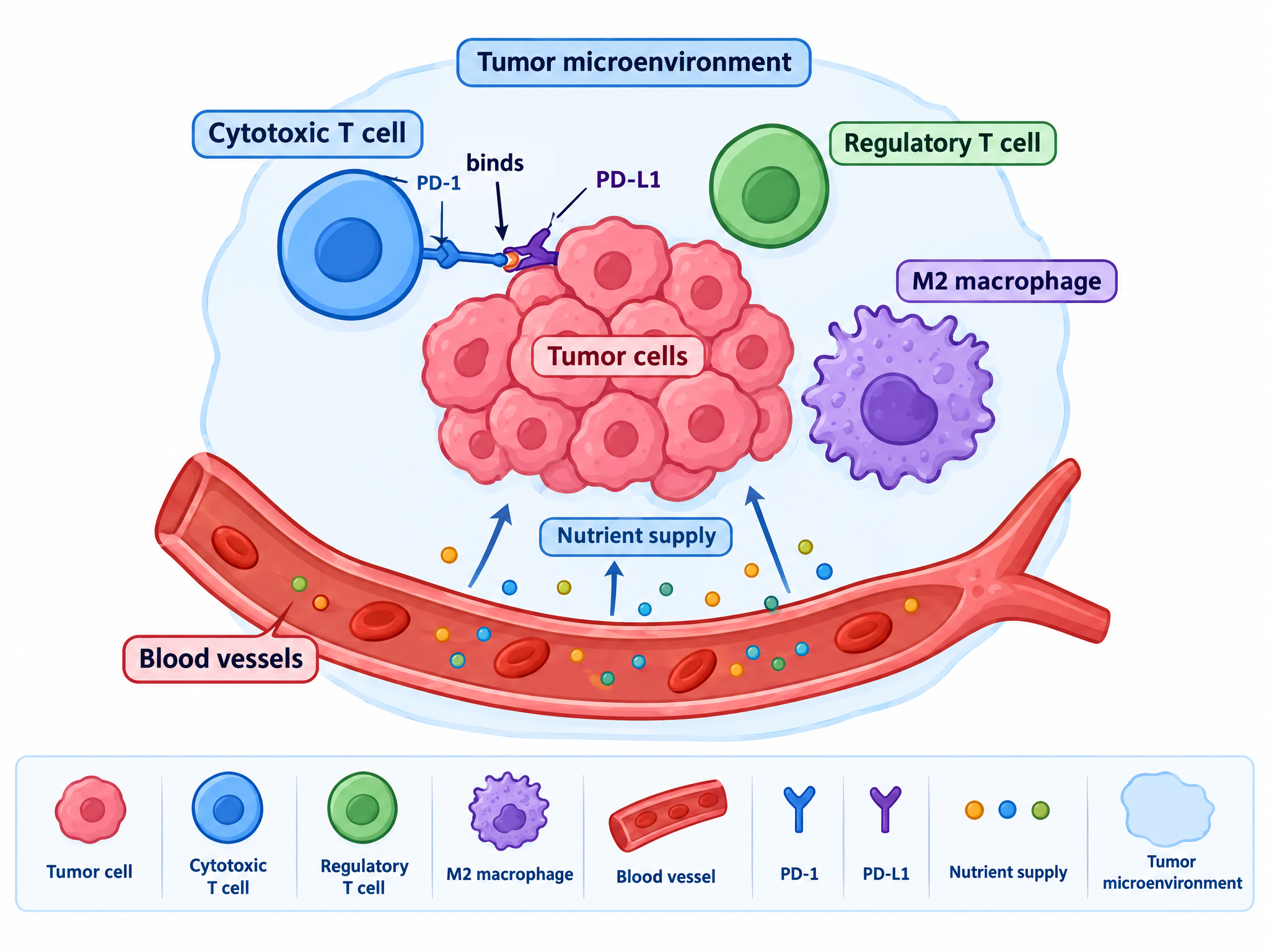
A tumor microenvironment review illustration: a cluster of tumor cells surrounded by cytotoxic T cells, regulatory T cells, M2 macrophages and blood vessels; show PD-1/PD-L1 checkpoint signals and nutrient supply from the vasculature. Balanced composition, labeled cell types.
Cos’è questo modello?
Review illustrations organize a whole research area into one scene — cell types, spatial relationships and signaling axes — giving readers a map before the text takes them through the details.
Dove si adatta
- Review article anchor figures
- Thesis introduction chapters
- Seminar background slides
- Grant proposal significance sections
Come renderlo tuo
Open the template in the studio
Generate the scene, then check which cell types and signals were included.
Rebalance the cast
Say "remove the macrophages and add fibroblasts" to match the compartment your review covers.
Highlight your axis
Ask to emphasize one pathway — e.g. "highlight the hypoxia axis from the vasculature" — while keeping the rest muted.
Domande frequenti
How dense can the scene get?
Keep it to five or six labeled elements per panel. If your review needs more, ask the editor to split the scene into two panels rather than crowding one.